Fertility Ultrasound
How are fertility scans performed?
To ensure we get the most accurate information, the Ultrasound Care team perform most fertility scans vaginally. Although some women experience a little discomfort, the scan should not be painful.
What is learned from a fertility scan?
The fertility assessment scan will provide your doctor information on the uterus, lining of the uterus, shape of the cavity and ovaries. Not all women are born the same, and ultrasound can help define if the uterus, Fallopian tubes and ovaries are present, fully formed and in the correct area. When assessing the uterus, the Ultrasound Care team obtains information about different shapes, problems with the muscle of the uterus, and problems with the lining which may affect your chances of conceiving and miscarriage.

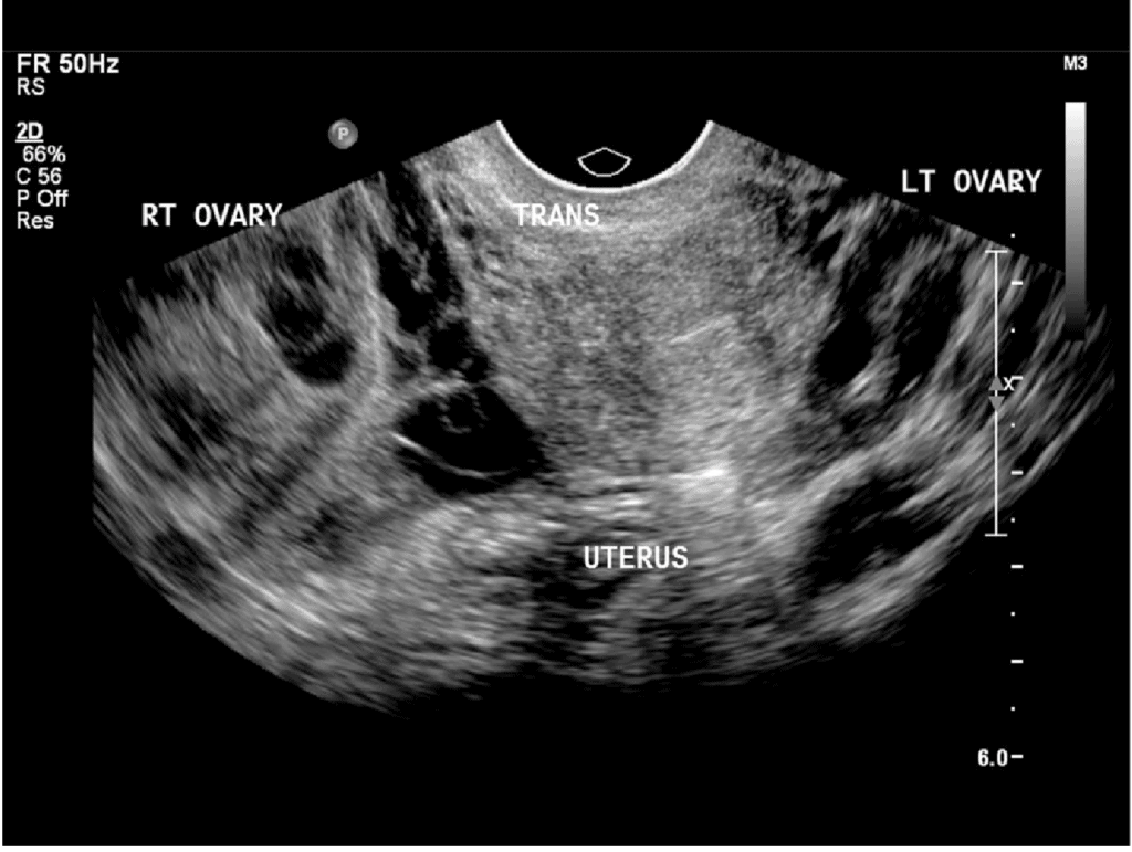
Pelvic and Ovary Ultrasound
What are the indications or reasons for having a pelvic and trans-vaginal ultrasound?
- Included as part of routine Well-Woman Screening
- Used to evaluate gynecological symptoms, including pelvic pain, irregular cycles, or other menstrual irregularities
- Helps confirm diagnoses such as uterine fibroids or ovarian cysts
- Assists in monitoring known conditions like fibroids or Polycystic Ovary Syndrome (PCOS)
- Supports fertility assessments or complements ongoing fertility treatments at other clinics
Pelvic Pain
Pain may be caused by any organ in the pelvis, and as such is often difficult to determine an exact cause without ultrasound. Discomfort with the onset of a period is normal, but severe pain should be investigated. It could be an indication of conditions such as endometriosis and cysts, these are best diagnosed by a Pelvic Ultrasound.
Abnormal Bleeding
Menstrual bleeding usually occurs every 28 days, although each cycle is different and some may be longer or shorter. At times the cycle may be irregular or bleeding may become heavy. This may be due to a temporary hormonal imbalance but it may also signal a problem in the uterus or ovaries such as fibroids or an endometrial polyp which is a small growth in the lining of the uterus. Polyps can cause heavier periods or unusual bleeding. Most polyps are not cancerous but may become so if not treated.
Fibroids
A fibroid is a benign growth of fibrous muscle tissue which develops in the wall of the uterus. Fibroids range in size from 5mm (the size of a pea) to 150mm (the size of a football). They are very common and are present in up to 30% of women. They generally do not cause any problems and many women go through life with their fibroids unnoticed.
Some women do however run into problems which may include:
- Infertility
- Heavy or irregular periods
- Pain
Fertility IVF Scan
Infertility may occur as a result of blockage of the fallopian tubes. Such a blockage may prevent the sperm from meeting the egg just before conception or prevent the embryo’s passage toward the uterus. Fertility may also be reduced if the fibroids significantly disrupt the cavity of the uterus.
Irregular bleeding can result due to enlargement or distortion of the uterine cavity by fibroids. A fibroid extending into the cavity can also cause heavy and irregular bleeding.
Follicular scans for IVF, also called a “baseline ultrasound,” is performed using a transvaginal ultrasound probe to examine the ovaries and uterus, to assess the number of antral follicles (small follicles containing eggs) within the ovaries, the thickness of the uterine lining, and check for any abnormalities that could affect implantation before starting IVF medication stimulation; this scan is crucial for determining the best treatment plan for the patient.
- Procedure:A transvaginal ultrasound probe is inserted into the vagina to get a clear image of the ovaries and uterus.
- What is assessed:
- Antral follicle count (AFC): The number of small follicles visible in the ovaries, which indicates ovarian reserve.
- Uterine lining thickness: Measuring the endometrium to ensure it’s thick enough for embryo implantation.
- Ovarian cysts: Checking for any cysts that may affect egg retrieval.
- Uterine fibroids: Identifying any fibroids that could interfere with implantation.
- Uterine shape and structure: Assessing for any abnormalities in the uterine cavity.
When is the scan done?- Baseline scan:This is usually performed on cycle day 2-5, right before starting IVF medications, to establish a starting point for monitoring follicle development.
- Monitoring scans:Throughout the stimulation phase of IVF, additional scans are done to track follicle growth and determine the optimal time for egg retrieval.
Obstetrical Anatomy Scan
During your second trimester, at 18 to 20 weeks pregnant, your doctor will request a 40-minute detailed obstetrical ultrasound. Sometimes called an anatomic ultrasound, this exam involves your sonographer taking many measurements of your baby from head-to-toe to determine how well your baby is growing. He or she will capture images to view the development of your baby’s brain, face, heart, spine, chest, major organs, arms, legs, feet, and hands. Your sonographer will also examine the position of your placenta, the vessels in the umbilical cord, the amount of amniotic fluid, and your cervix, uterus, ovaries, and bladder for abnormalities. It’s also during this exam when the gender of your baby may first be visible – if the baby is in a good position.