Most people assume that heart problems announce themselves loudly, a dramatic episode of chest pain, a sudden collapse, something that leaves no doubt that an emergency is happening. But that's not how it works for most people. Many of the most serious heart conditions develop slowly and silently, often over months or years, long before …
Most people assume that heart problems announce themselves loudly, a dramatic episode of chest pain, a sudden collapse, something that leaves no doubt that an emergency is happening.
But that’s not how it works for most people.
Many of the most serious heart conditions develop slowly and silently, often over months or years, long before any noticeable symptoms appear. By the time something feels wrong, the underlying issue may have been progressing for a long time.
An echocardiogram, commonly called a heart ultrasound or simply an “echo,” is one of the most valuable tools available for understanding what’s actually happening inside your heart, often well before a problem becomes serious.
What Is an Echocardiogram and How Does It Work?
An echocardiogram uses high-frequency sound waves to create real-time, moving images of the heart. Unlike many other types of cardiac testing, an echo allows a sonographer and reviewing physician to actually watch the heart in motion, the chamber walls contracting and relaxing, the four valves opening and closing with each heartbeat, and blood flowing through the heart’s structures.
This sets it apart from other common cardiac tests. A chest X-ray provides a static image showing the heart’s general size and shape, but no information about how it’s functioning. An EKG (electrocardiogram) records the heart’s electrical activity, useful for detecting rhythm abnormalities, but it doesn’t show the physical structure of the heart itself.
An echocardiogram bridges that gap, offering a detailed, dynamic view of both structure and function in a single, non-invasive test.
What an Echocardiogram Can Detect
Because an echo provides a live view of the heart, it can identify a wide range of conditions, including:
Heart muscle abnormalities, including areas where the muscle isn’t contracting properly due to reduced blood flow, which can be an early indicator of coronary artery disease.
Valve problems, such as a valve that isn’t opening fully (stenosis) or one that’s allowing blood to leak backward (regurgitation). The heart has four valves, and dysfunction in any of them can place additional strain on the heart over time.
Fluid buildup around the heart (pericardial effusion), which can occur due to inflammation, infection, or other underlying conditions.
Chamber enlargement or thickening, which often develops as a response to high blood pressure or other conditions that force the heart to work harder than it should.
Overall pumping efficiency, often measured as “ejection fraction,”, a key indicator of how well the heart is functioning as a pump.
Who Should Consider Getting an Echocardiogram?
A common misconception is that cardiac imaging is only for people who have already experienced a heart attack or have been diagnosed with heart disease. In reality, there are many situations where getting an echocardiogram proactively makes sense, and where catching an issue early can make a significant difference.
Unexplained shortness of breath. If you’ve noticed that you’re more breathless than usual, especially when lying down, during light activity, or for no clear reason, an echo can help determine whether the heart is contributing to this symptom.
Heart palpitations or irregular heartbeat. A fluttering, racing, or skipping sensation in the chest can have many causes, but an echo can help identify whether there’s an underlying structural issue contributing to it.
Difficult-to-control high blood pressure. Chronic high blood pressure can cause the heart muscle to thicken over time (a condition called left ventricular hypertrophy). An echo can detect this early, before it progresses further.
A history of heart murmur. If you’ve ever been told you have a heart murmur, even if it was mentioned casually years ago, an echo can clarify whether it’s related to a valve issue that should be monitored.
Family history or risk factors. A family history of heart disease, a personal history of diabetes, or other cardiovascular risk factors are all good reasons to consider establishing a baseline.
Age-related screening. For adults over 50 who have never had any form of cardiac imaging, a baseline echocardiogram provides a valuable reference point for future comparisons and offers either reassurance or early detection.
What Happens During an Echocardiogram Appointment
One of the most reassuring things about an echocardiogram is how simple and comfortable the experience is.
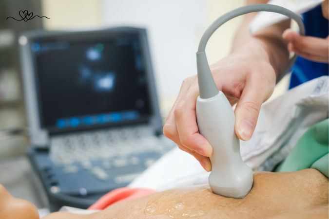
The appointment typically takes between 45 and 60 minutes. You’ll lie on an exam table, usually positioned on your left side, which helps bring the heart closer to the chest wall for clearer imaging. A small amount of gel is applied to the chest, and the sonographer uses a handheld transducer to capture images from several different angles.
During the scan, you may be asked to breathe in a certain way, exhale, or briefly hold your breath. These instructions help reduce motion and allow the sonographer to capture the clearest possible images of the heart’s structures.
A standard resting echocardiogram involves no radiation, no injections, and no special preparation. You can eat and drink normally beforehand, and resume your normal activities immediately afterward.
Once the images are captured, they are reviewed by a board-certified radiologist or cardiologist, who prepares a detailed report outlining the findings.
Understanding the Cost Difference
Cardiac imaging can come with a significant price tag, particularly at hospital-based facilities. A hospital echocardiogram can cost well over a thousand dollars for patients without insurance, and even insured patients can face substantial out-of-pocket costs due to deductibles, copays, and coinsurance.
At a self-pay imaging clinic, the same type of scan, performed by a qualified cardiac sonographer and reviewed by a board-certified radiologist, is often available at a significantly reduced price.
This matters because cost is consistently cited as one of the leading reasons people delay or avoid diagnostic imaging altogether. And when it comes to cardiovascular health, the leading cause of death among adults in the United States, delaying a simple, painless scan is a risk that’s worth reconsidering.
Why Early Information Matters
The heart works continuously, beating roughly 100,000 times per day, every day of your life, without pause.
Getting an echocardiogram isn’t something to approach with anxiety; it’s an opportunity to gain clarity. If the results show a healthy heart, that’s a valuable peace of mind. And if something does need attention, identifying it early, while there’s still time to address it through lifestyle changes, monitoring, or treatment, is one of the most proactive steps you can take for your long-term health.
Rather than waiting for symptoms to force a conversation about your heart health, an echocardiogram offers a way to start that conversation on your own terms, while you still have the most options available to you.
Take the Next Step Toward Understanding Your Heart Health
If any of the situations described above sound familiar, or if you simply want a clearer picture of how your heart is functioning, scheduling an echocardiogram is a straightforward, accessible step. At a dedicated imaging clinic offering self-pay options, you can receive a thorough, professionally reviewed scan without the long wait times or high costs often associated with hospital-based testing.
Understanding your heart doesn’t have to wait for a crisis. It can start with a simple, painless scan today.
Sign up for free Session!
It’s easy and free!